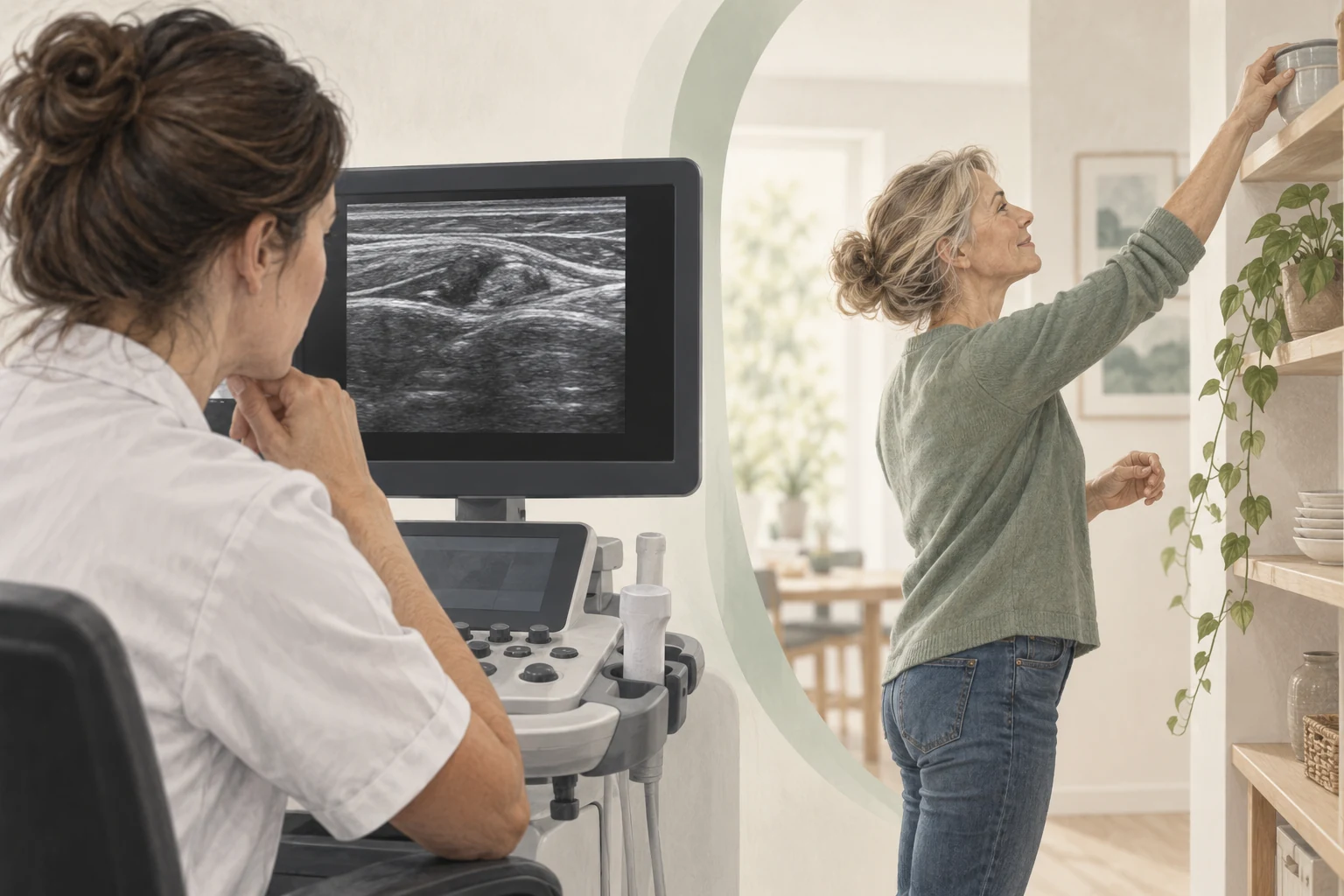
An ultrasound starts with a clinical question
Before an ultrasound head is placed on the skin, it should be clear what one wants to know. Is there any doubt about the structure involved, is the course not appropriate for the probable diagnosis or is additional information necessary for a treatment? Those are targeted research questions.
A general desire to see what is wrong is understandable, but too broad. An ultrasound shows certain structures and cannot show all the causes of pain. The question in advance therefore also determines how useful the result will be.
Ask your clinician what's going to be different in a normal and abnormal rash. If both answers produce exactly the same treatment plan, the added value of the study may be limited.
What can ultrasound show?
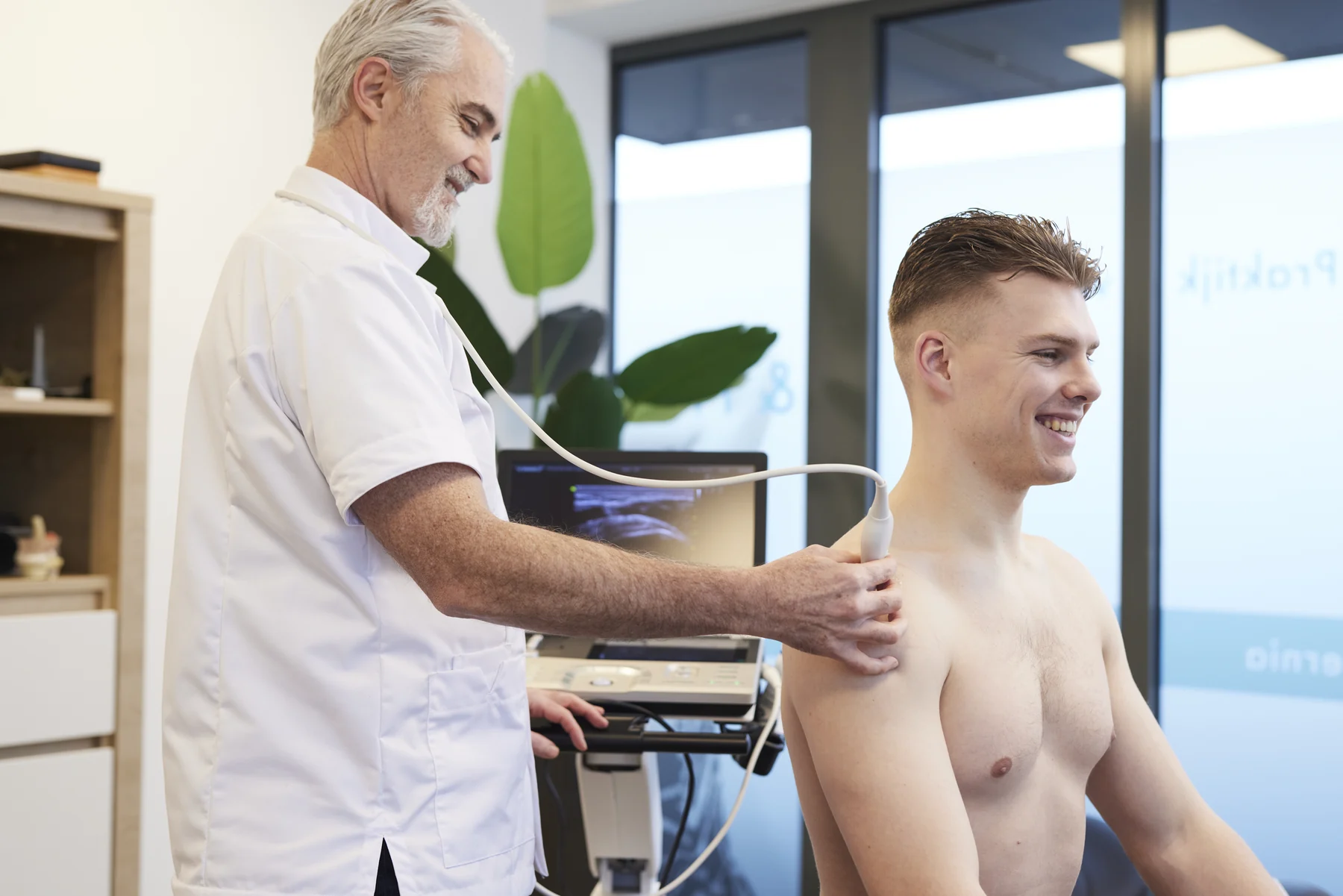
In ultrasound, sound waves are used to make images. Treads and surrounding tissues can be viewed. The researcher can, among other things, look at form, structure and certain changes in the area.
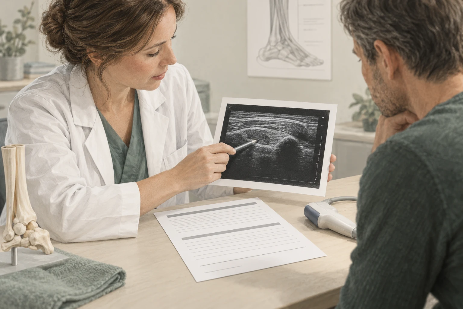
However, an image is not a direct pain meter. A thickening or other finding should match the location and pattern of your symptoms. The words in a report describe what is visible; the meaning for your functioning requires interpretation.
The information from Thuisarts about shoulder problems explains why a found particularity is not automatically the cause of the pain. That is an important starting point when you look at the images together.
When can research be sufficient without ultrasound?
Some symptoms of the tendons can be sufficiently likely to be detected with a recognizable story and physical examination. The Dutch Achilles guideline does not recommend additional imaging in an appropriate set of clinical criteria.
That doesn't mean the clinician only works on feeling. The site of pain, relationship with load and findings in studies are assessed together. This creates a well-founded work diagnosis.
No ultrasound can therefore be a substantive choice. Question on which the diagnosis is based and when is re-evaluated. A clear plan gives more certainty than a scan without explaining what the findings mean.
When does an ultrasound become more interesting?
In case of diagnostic doubt or an unexpected course, imaging can add more. A new change after trauma may also raise another research question. The right route depends on the situation; sometimes a different form of research is needed.
The moment an ultrasound is considered is not the same for all tendons. In one symptom clinical research is clear, in another there is doubt about multiple structures. Therefore, the duration of symptoms alone does not automatically determine the necessity.
Describe what's changed in a follow-up appointment. It's in another place now or I'm suddenly losing power helps more than just saying that the symptom still exists. This allows the clinic to determine whether the original question should be reviewed.
How do you discuss the results?
A good result discussion distinguishes between three layers: what has been seen, how certain is the interpretation and what changes this plan? Ask for all three. This way you prevent a technical word unintentionally from lingering like a serious diagnosis.
For example, use these questions:
- Does this finding fit in with my symptoms and investigations?
- Does this happen in people with no pain?
- Does this change my treatment or build-up?
- What uncertainty remains?
A normal ultrasound doesn't mean your symptoms aren't real. The meaning of a normal result depends on the original question. Sometimes a probable explanation is less likely, while other possibilities still need to be considered.
ultrasound within an intake
At Shockwave Clinics the intake is paid and ultrasound is free when it is clinically meaningful. That offer does not change the substantive balance: imaging should have a purpose. It's not a mandatory part for everyone.
Bring earlier images or reports when you have them. A repetition can sometimes be useful, but earlier research can also provide sufficient information. It prevents the same question from being re-examined without anything changing.
After the intake, you especially want an understandable answer to the question of how to proceed. An ultrasound can support that answer. The research has been successful when it helps make the right decision, not only when a striking picture is found.
Frequently asked questions
Can an ultrasound prove where my pain comes from?
An ultrasound can support a suspected statement or make it less likely. The images must be placed next to the symptoms pattern and physical examination. A visible anomaly alone does not automatically prove that it is the complete cause of your pain.
Isn't an ultrasound a less careful intake?
No. If story and research are sufficiently clear and imaging does not change the plan, it can be careful to refrain from an ultrasound. However, it is necessary to explain the basis of the diagnosis and where re-evaluation or additional studies may be necessary.
Can I get a shockwave right away if the ultrasound shows anything?
A finding is not an automatic treatment indication. First, it must be clear whether it fits the symptoms and which approach is sufficiently substantiated. Sometimes the result changes the choice precisely in the direction of another treatment or further assessment.
Is the intake free if you don't need an ultrasound?
No. The intake at Shockwave Clinics has been paid. Ultrasound is free when it is clinically meaningful. The consultation also includes assessment and discussion of the follow-up; the making or not making an ultrasound does not change the intake into a free appointment.
Sources and evidence
- guideline Achilles tendinopathy – diagnosis (2020) ↗
Clinical diagnosis, targeted imaging and limits of ultrasound information.
- Thuisarts – My shoulder hurts. ↗
Anamnesia and physical examination; imaging is usually not necessary in case of short-standing shoulder problems.
A personal follow-up step
From insight
to an appropriate plan.
At a paid intake, we'll discuss your symptom and do targeted research. ultrasound is included at no extra cost when it makes sense. Any treatments are agreed separately.
View the initial consultation