Read the question and conclusion first
A report usually starts from a research question and ends with a conclusion. Those parts help put the technical description in between. A loose term from the middle of the text can sound much heavier than the overall assessment.
Also check which body part and which side have been examined. When multiple structures are mentioned, not every detail is automatically the explanation for your symptom. The report may contain relevant and additional findings.
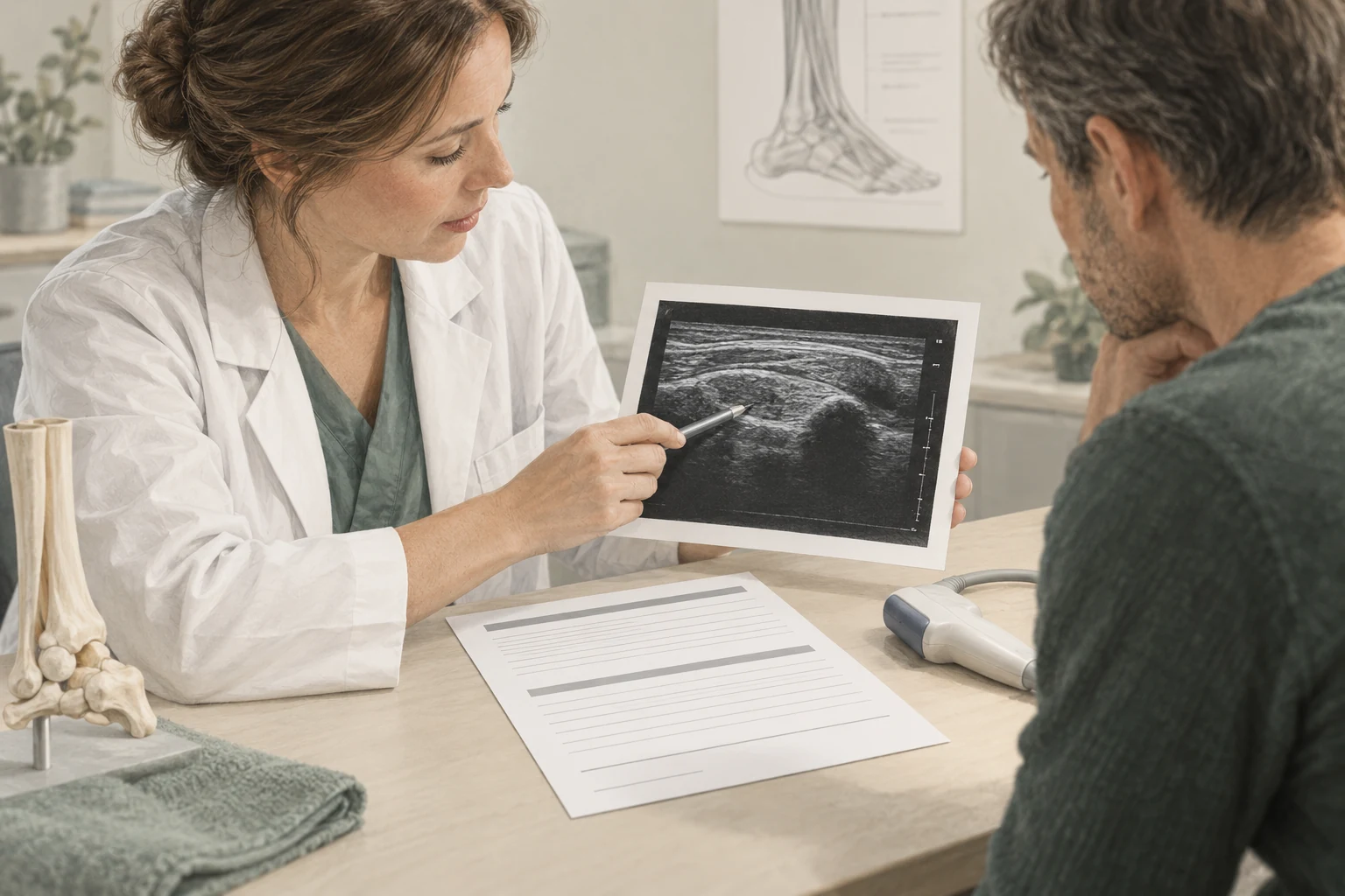
Ask the practitioner to summarise the conclusion in plain language. A useful answer indicates what probably has meaning, which remains uncertain and what follow-up results.
What do thickness and structure mean?
A thickened tendon is a description of the shape. A modified structure means that the image deviates from the usual pattern. The Achilles guideline mentions thickness and structure as components of reporting.
These descriptions are not the same as a percentage that the tendon can still bear. From a millimetre you cannot independently deduce how much you can walk or lift. Also, a difference with another measurement is not automatically a significant change.
The interpretation depends on location, technique and clinical situation. Ask not only whether the tendon is thick, but whether that finding fits the grievance pattern and has consequences for the accompaniment.
Echoarm, Doppler and vascularization
Echoarm describes how an area appears on the image. It's a technical description, not a diagnosis in itself. The investigator shall assess the pattern and its consistency with other characteristics.
Doppler is used to make signals of blood flow visible. Terms such as vascularization or Doppler signal refer to that assessment. You cannot simply deduce that a tendon is actively broken or that extra blood flow is always good or bad.
In such words, ask what clinical significance is assigned to it. When a term is mainly registered for completeness, it does not need to become the center of your recovery plan. The description and treatment advice remain separate steps.
Calcination and tear require specific explanation
Calcination means that lime-containing material is visible. The significance varies from one person to another. A lime finding is not automatically the only cause of pain or a general indication of how to treat it with a device.
In the event of a rupture, it is relevant, inter alia, whether it is a partial or complete interruption, how the symptom arose and what function is present. Therefore, do not read the word separately from the conclusion and physical examination.
These terms deserve explanation, without immediately minimising or dramatizing them. A random finding and an acute change with loss of strength pose very different questions. The report should be discussed in this context.
A picture doesn't just predict the course
The Dutch guideline on Prognosis in Achilles Tendon symptoms advises not to use imaging to predict the course of symptoms. The severity of the image is therefore not a simple calendar for recovery.
That is relevant when you see words as degenerative or chronic. They don't automatically mean that improvement is impossible. Ask which information is used to track the course of events, for example symptoms about physical load and functioning.
Therefore, treatment does not need to be continued until every detail looks normal on the ultrasound. The reason for further care should be in line with your clinical situation and goals, not just the desire to get a report without any different terms.
Make the report a useful conversation
You can add the following note to the report:
| In the report | My question |
|---|---|
| Technical finding | What does this mean in plain language? |
| Probable diagnosis | Does this fit in with my symptoms and investigations? |
| Insecure formulation | What information is still missing? |
| Opinion | What does this change my daily plan? |
Keep the original report and a brief summary of the explanation. Next time you meet, you're gonna avoid remembering just one disturbing word.
If different carers explain the outcome differently, ask which part leads to the difference. Sometimes it is the same finding with a different assessment of its meaning. A targeted conversation about this is more useful than constantly scanning without a clear question.
Frequently asked questions
Does degenerative mean my tendon is worn out and irreparable?
Not automatically. The word describes an interpretation of tissue change and does not in itself say enough about your functioning or recovery possibilities. Ask what practical significance it has in your report and on which the treatment advice is based.
Is a thicker tendon always a worse tendon?
No. Thickness is one characteristic and must be read in context. You can't distract yourself from strength, pain or recovery time. The place, other findings, the physical examination and your possibilities in daily life count together.
Does more Doppler signal mean I can't practice?
That doesn't automatically follow from such a finding. The exercise advice is determined by the clinical situation and the overall plan. Ask what meaning the clinician gives to the signal and what response to load you should follow in your situation.
Why does it say "probably" or "appropriate"?
Imaging doesn't always give absolute certainty. Such formulations reveal how the investigator interprets the finding. Ask which alternatives are still possible and whether that uncertainty affects treatment or the need for additional research.
Sources and evidence
- guideline Achilles tendinopathy – diagnostic image finding (2020) ↗
Reporting of tendon thickness, structure, vascularisation and calcifications.
- guideline Achilles tendinopathy – prognostic factors on imaging (2020) ↗
Do not use imaging to predict the course of symptoms.
A personal follow-up step
From insight
to an appropriate plan.
At a paid intake, we'll discuss your symptom and do targeted research. ultrasound is included at no extra cost when it makes sense. Any treatments are agreed separately.
View the initial consultation