The need for a visible recovery certificate
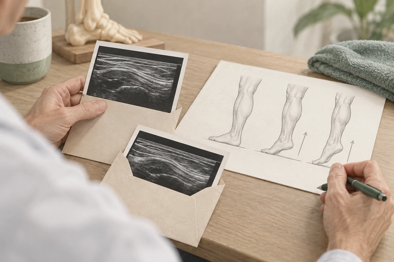
If you've had symptoms for a long time, it's understandable that you want to see on an image that tendon gets better. A new ultrasound then seems to be an objective check. However, the question of whether a tendon looks different is not the same as whether you work better.
You can do more while certain image features remain present. Also, a measurement can change without significantly improving your daily life. It is therefore important to determine in advance which question the repeat ultrasound should answer.
So don't just ask if the tendon looks better, but what changes that information about the plan. A scan without a decision question can promise reassurance that the result will not give.
What do you advise guidelines about following?
The Dutch Achilles guideline discusses measuring instruments for the treatment course. It focuses on symptoms, functioning and personal goals. Routine imaging does not automatically add value to that evaluation.
The individual module on prognostic image characteristics advises no imaging to predict the course of symptoms. So a scan cannot reliably give your personal end date of recovery.
These are advice for Achilles Tendon charges. The details may differ with other diagnoses. The broader principle remains that diagnostics, prognosis and evaluation are different questions and are not automatically answered with the same research.
What better to follow in your daily life?
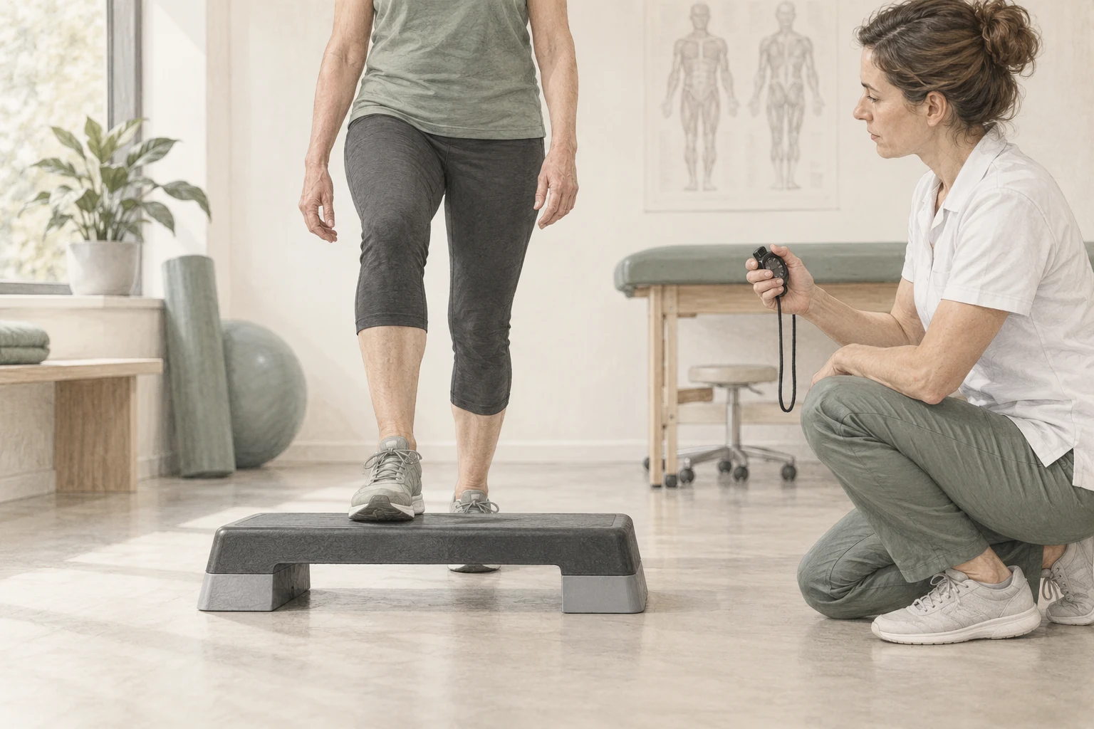
Choose an activity that matches your goal. Write down how much you do, how that goes, and what reaction follows later. That makes it visible whether your possibilities are really changing.
With a hiking destination, the duration or number of breaks can be relevant. A shoulder target may be a recurring reach or tilt task. Compare as many similar circumstances as possible. A quiet holiday and a heavy working week give a distorted image.
A questionnaire or functional test may supplement the assessment. Ask what the instrument measures and how the outcome is used. A test is a tool for decision-making, not a single digit that summarises your entire recovery.
When can a new ultrasound make sense?
An unexpected change may raise a new diagnostic question. Think of another pain site, a new incident or a sudden loss of function. Then the ultrasound isn't just about controlling the original finding.
A change in treatment choice may also require additional information. It is therefore important that the controller explains what decision it depends on. Repeating the same scan because a few weeks has passed, is another reason than targeted a new question investigations.
Bring earlier reports. This will enable us to look at what has already been established and what is still missing. Sometimes revisiting or discussing existing information is sufficient; sometimes new imaging is appropriate.
Compare measurements carefully
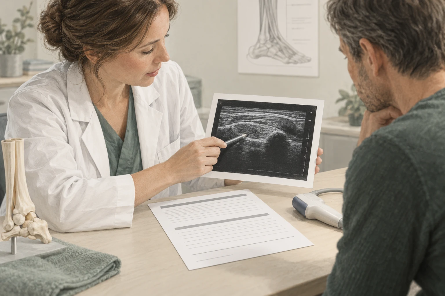
A difference between two echoes requires interpretation. The place of measurement, execution and description used may affect what is in a report. A few other words do not automatically mean that the situation has changed greatly.
Ask if the studies are sufficiently comparable. Was the same part viewed, the same finding measured, and is the difference big enough to have meaning? Those questions are part of the expert assessment.
A real difference in structure has yet to be linked to your clinical development. The image can provide additional information, but is not an independent report rate for your commitment or for the quality of the treatment.
A decision tree for the conversation
You can make a proposal for repeatechography along four steps:
- Is there a new question? Name what has become unclear since the previous investigation.
- Can ultrasound answer that question? Sometimes a different study or clinical assessment fits better.
- Is the outcome changing the plan? Discuss what a possible outcome means.
- What's next? Keep functioning and goals visible.
If the main reason is reassurance, discuss what security you're looking for. Maybe you want to know if you can build. That question is often better answered with clinical information and an appropriate load plan than with just an image.
Therefore, following recovery does not have to be less careful without repeated scans. Carefulness means that you collect the information needed for the next decision and that new signals are seriously assessed.
Frequently asked questions
Does my ultrasound have to be normal before I can exercise again?
That's not a general condition. Returning to sport requires assessment of symptoms, function and the burden the sport imposes. The role of imaging depends on the diagnosis. Ask which criteria are used in your situation and why.
Does a better ultrasound prove that shockwave has worked?
Not on its own. A change in image characteristics is a different outcome than an improvement in pain and functioning. In addition, several parts of the treatment plan may have an impact. A personal evaluation should therefore be broader than comparing two images.
What if I function better but the ultrasound is the same?
That could happen. Discuss which finding is still visible and whether it has consequences. An unchanged image doesn't have to mean that your clinical progress is unimportant. The treatment advice is part of the overall pattern and your goals.
When do I ask for a new assessment?
In case of an unexpected change, a new injury moment or persistent doubt about the diagnosis, consultation is useful. The operator then determines whether physical examination, imaging or referral is necessary. A new assessment does not necessarily mean a new ultrasound.
Sources and evidence
- guideline Achilles tendinopathy – measuring instruments (2020) ↗
Clinical monitoring of symptoms and functioning; no routine imaging for evaluation.
- guideline Achilles tendinopathy – prognostic factors on imaging (2020) ↗
Do not use imaging to predict the course of symptoms.
A personal follow-up step
From insight
to an appropriate plan.
At a paid intake, we'll discuss your symptom and do targeted research. ultrasound is included at no extra cost when it makes sense. Any treatments are agreed separately.
View the initial consultation